Chest Muscles Diagram - Chest Muscle Anatomy Anatomy Drawing Diagram : The dominant muscle in the upper chest is the pectoralis major.. The word sternum originates from the ancient greek word 'sternon', meaning chest. Jan 04, 2018 · doing leg exercises at home is probably a lot easier than you realize. The pectoralis major, pectoralis minor, serratus anterior and subclavius. Learn vocabulary, terms, and more with flashcards, games, and other study tools. Set a bench to a 45 degree incline.
Start studying 14.4 chest muscles. (1) the pectoralis major, and (2) the pectoralis minor. The sternum is also known as the breastbone. It spreads out like a fan and covers the rib cage like an armor plate. It is a flat bone that articulates with the clavicle and the costal cartilages of the upper 7 ribs (true ribs), while the 8th, 9th and 10th ribs (false ribs) are indirectly attached with sternum via costal cartilage of the ribs above.
Chest Muscles Anatomy Images Stock Photos Vectors Shutterstock from image.shutterstock.com The chest muscles are made up of the pectoralis major and, underneath that, the pectoralis minor. The sternum is located along the body's midline in the anterior thoracic region just deep to the skin. Your pectoralis major and pectoralis minor muscles make up most of the muscle mass in your chest. The pec major) is the one that commands the most real estate. Related posts of chest muscles diagram muscle gross anatomy. I often get asked, how can i build thick powerful pecs? Learn vocabulary, terms, and more with flashcards, games, and other study tools. Jan 04, 2018 · doing leg exercises at home is probably a lot easier than you realize.
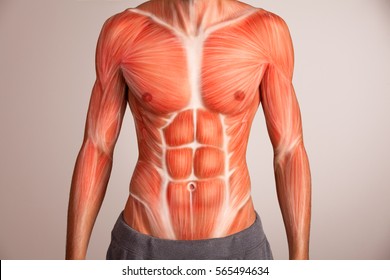
Your pectoralis major and pectoralis minor muscles make up most of the muscle mass in your chest.
The chest is part of a larger group of pushing muscles found in the upper body. Chest muscles, chest muscle diagram. It contains organs including the heart lungs and thymus gland as well as muscles and various other internal structures. Enter the answer length or the answer pattern to get better results. Chest muscles, chest muscle diagram. Related posts of chest muscles diagram muscle gross anatomy. The sternum is also known as the breastbone. This is a table of skeletal muscles of the human anatomy. (1) the pectoralis major, and (2) the pectoralis minor. The primary function is certainly to provide support to the skeletal system and to facilitate its movements. It's important to note that it attaches to your upper arm. Well now we have the answers for you, cep training revolutionary muscle growth secrets <== click here below is a diagram showing the chest muscles depicting where the different exercises target. There are red muscles stretched over the stomach, chest, and shoulders, and on top of each breast is a complicated structure made out of milk ducts, which appears in pieces fanned out that make it.
This is a table of skeletal muscles of the human anatomy. The sternum is located along the body's midline in the anterior thoracic region just deep to the skin. Jan 04, 2018 · doing leg exercises at home is probably a lot easier than you realize. Below is a diagram showing the chest muscles depicting where the different exercises target. It contains organs including the heart lungs and thymus gland as well as muscles and various other internal structures.
Chest Muscles Anatomy Images Stock Photos Vectors Shutterstock from image.shutterstock.com The chest muscles are made up of the pectoralis major and, underneath that, the pectoralis minor. The dominant muscle in the upper chest is the pectoralis major. Related posts of chest muscles diagram muscle gross anatomy. Well now we have the answers for you, cep training revolutionary muscle growth secrets <== click here below is a diagram showing the chest muscles depicting where the different exercises target. Related posts of muscle diagram for chest and back back muscles anatomy video. The major muscle in the chest is the pectoralis major. There are multiple functions of these chest. Human muscle diagram graph diagram.
The chest, as part of this group, enables you to perform pushing actions such as the barbell bench press or a daily activity such as moving a heavy dresser.
Surgical anatomy of the chest wall thoracic key. If you are pulling your muscles more than enough and doing burdening lots of effort, then the pain that is going to felt by you is no the symptom of the muscular chest pain. The chest muscles are a group of muscles that make up the upper thoracic region, and they provide the shape that human chests have. Human muscle diagram graph diagram. The chest muscles the 2 pectoralis muscles, the pectoralis major and the pectoralis minor (the larger and smaller muscles of the chest) connect the front of the chest wall with the humerus (upper arm bone) and shoulder (fig). This is a table of skeletal muscles of the human anatomy. It also protects several vital organs of the chest, such as the heart, aorta, vena cava, and thymus gland that are located just deep to the sternum. It's the large muscle that covers the upper chest area, starting at the sternum and collar bone and from there attaches to your upper arm. The major muscle in the chest is the pectoralis major. Muscle gross anatomy 12 photos of the muscle gross anatomy gross anatomy of cardiac muscle, gross anatomy of skeletal muscle worksheet, gross muscle anatomy test, muscle gross anatomy quiz, muscular system gross anatomy chapter 10, human muscles, gross anatomy of cardiac muscle, gross anatomy of skeletal muscle worksheet, gross. The dominant muscle in the upper chest is the pectoralis major. It's important to note that it attaches to your upper arm. Related posts of chest muscles diagram muscle gross anatomy.
The chest muscles of our body can get pulled and strained. Chest muscles anatomy (1) pectoralis major muscle. Of the two chest muscles, the pectoralis major (a.k.a. The chest is part of a larger group of pushing muscles found in the upper body. There are red muscles stretched over the stomach, chest, and shoulders, and on top of each breast is a complicated structure made out of milk ducts, which appears in pieces fanned out that make it.
Female Chest Muscles Anatomy Diagram Function Body Maps from post.healthline.com The primary function is certainly to provide support to the skeletal system and to facilitate its movements. It contains organs including the heart lungs and thymus gland as well as muscles and various other internal structures. Muscle gross anatomy 12 photos of the muscle gross anatomy gross anatomy of cardiac muscle, gross anatomy of skeletal muscle worksheet, gross muscle anatomy test, muscle gross anatomy quiz, muscular system gross anatomy chapter 10, human muscles, gross anatomy of cardiac muscle, gross anatomy of skeletal muscle worksheet, gross. Muscles allow a person to move, speak, and chew. Related posts of muscle diagram for chest and back back muscles anatomy video. I often get asked, how can i build thick powerful pecs? Whenever you press something in front of you, they work, for example, when you do barbell or dumbbell bench presses. Of the two chest muscles, the pectoralis major (a.k.a.
It contains four muscles that exert a force on the upper limb:
If a pair is in your gym bag on chest day, your muscles and joints will both thank you. Unfortunately, in many cases, that's as far as the doctor takes the diagnosis. The major muscle in the chest is the pectoralis major. To fully develop your chest, you need to hit it with heavy weight using a couple smartly chosen exercises. Whenever you press something in front of you, they work, for example, when you do barbell or dumbbell bench presses. If you are pulling your muscles more than enough and doing burdening lots of effort, then the pain that is going to felt by you is no the symptom of the muscular chest pain. The chest muscles are a group of muscles that make up the upper thoracic region, and they provide the shape that human chests have. The usual cause of overstretched chest muscles can be the over exercising. The primary function is certainly to provide support to the skeletal system and to facilitate its movements. Related posts of muscle diagram for chest and back back muscles anatomy video. Chest muscles, chest muscle diagram. It is a flat bone that articulates with the clavicle and the costal cartilages of the upper 7 ribs (true ribs), while the 8th, 9th and 10th ribs (false ribs) are indirectly attached with sternum via costal cartilage of the ribs above. Chest wall pain is caused by problems affecting the muscles, bones and/or nerves of the chest wall.


0 Komentar